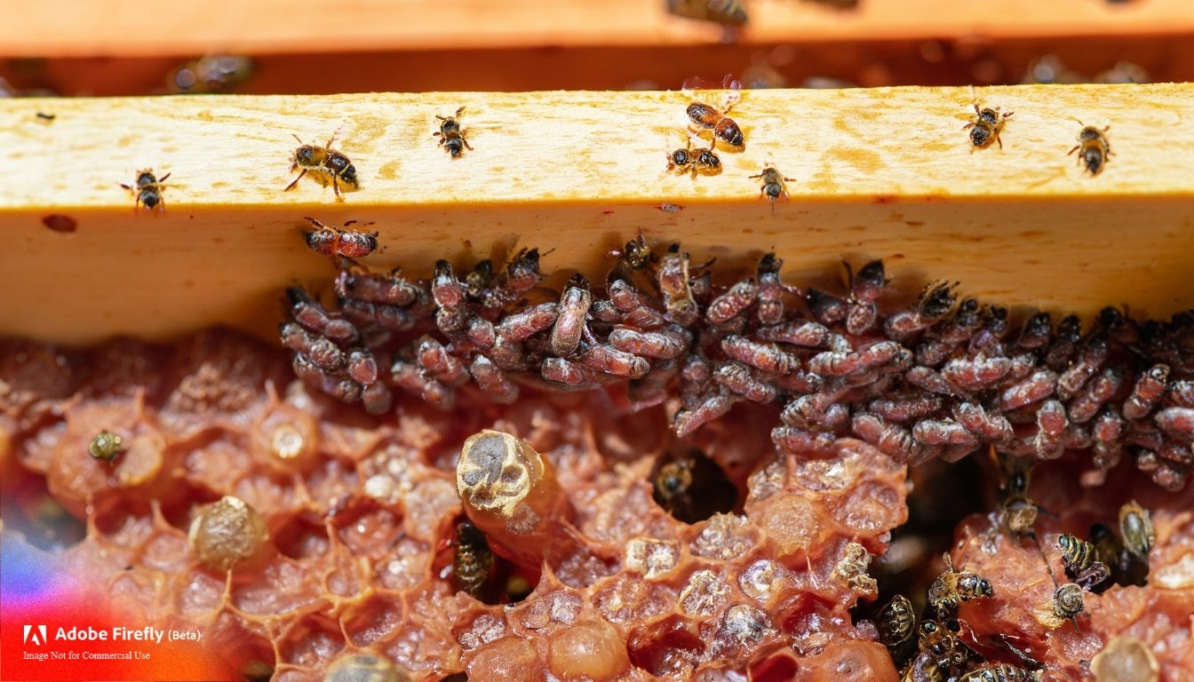
Varroa mites (Varroa destructor) are parasitic mites that pose a significant threat to honeybee colonies worldwide. These tiny arachnids attach themselves to honeybees and their developing brood, feeding on their bodily fluids and transmitting harmful viruses. Recognizing and understanding Varroa mites’ appearance is crucial for beekeepers to detect infestations and implement timely control measures. In this comprehensive guide, we’ll explore the physical characteristics of Varroa mites, their life cycle, and the implications of their presence in bee colonies.
Post Contents
The Anatomy of Varroa Mites
Varroa mites are minute creatures, barely visible to the naked eye. They belong to the family Varroidae, and their body structure consists of several distinct parts:
- Legs: Varroa mites possess eight legs, a defining characteristic of arachnids. These legs are adapted for clinging to honeybees and crawling among the comb cells.
- Mouthparts: The mouthparts of Varroa mites are designed for piercing the cuticle of honeybees and feeding on their hemolymph (blood equivalent). These mouthparts allow the mites to extract nutrients from their hosts.
- Body: The mites have a flattened, oval-shaped body with a hard exoskeleton. This exoskeleton provides protection and helps them withstand the rigors of their environment.
- Color: Varroa mites can vary in color, ranging from light brown to reddish-brown. Their coloration often depends on their age and the specific stage of their life cycle.
Life Cycle of Varroa Mites
To better understand the appearance of Varroa mites, it’s essential to grasp their life cycle. The life cycle of Varroa mites consists of several stages:
- Phoretic Phase: Adult female Varroa mites attach themselves to adult honeybees and are transported into the beehive. During this phase, they feed on the bee’s hemolymph and continue to reproduce.
- Reproductive Phase: The female mites enter brood cells that contain developing bee larvae. They lay eggs on the bee larvae, and once the cell is capped, the mites begin reproducing. The eggs hatch into protonymphs, which then develop into deutonymphs and adult male and female mites.
- Offspring Development: As the bee larvae develop, the mites continue to reproduce inside the capped cells. Female mites lay eggs on both male and female bee larvae.
- Emergence: Once the bee larvae pupate and emerge as adult bees, the mites also emerge from the cells. The mites that were reproducing inside the cells are now ready to infest other bees and continue their life cycle.

Identifying Varroa Mites
Given their small size, identifying Varroa mites requires careful observation and, ideally, using magnifying tools. Beekeepers commonly employ the following methods to detect Varroa mites:
- Sticky Boards: Placing sticky boards underneath beehives can capture mites that fall off bees. By counting the number of mites on the sticky board over a period, beekeepers can estimate the infestation level.
- Alcohol Wash: Beekeepers can take a sample of adult bees from a hive and wash them in alcohol. The alcohol dislodges the mites from the bees, allowing beekeepers to quantify the mite load.
- Powdered Sugar Roll: This method involves collecting a sample of bees and rolling them in powdered sugar. The sugar dislodges the mites, which can be counted after falling through a mesh screen.
- Visual Inspection: Careful inspection of adult bees, brood cells, and drone pupae can reveal the presence of mites attached to their bodies.
What to Look for: Visual inspection for Varroa Mites
When conducting a visual inspection, beekeepers should focus on specific areas where Varroa mites are commonly found:
- Adult Bees: Look for mites attached to the body of adult bees, particularly in the joints and between body segments. Mites may appear as small, oval-shaped creatures.
- Brood Cells: Inspect capped brood cells for mites that may be visible through the cell cappings. Mites may be found on the bees emerging from the cells.
- Drone Brood: Varroa mites often prefer drone brood cells for reproduction. Check drone pupae for mites nestled between the abdominal segments.
- Mite-Infested Bees: Bees infested with Varroa mites may display signs of deformities, weakened wings, or other physical abnormalities.
Implications of Varroa Mite Infestations
Recognizing Varroa mite infestations is crucial because these parasites can devastate honeybee colonies. Here are some of the potential implications of unchecked Varroa mite populations:
- Weakened Colonies: High Varroa mite infestations can weaken bee colonies, reducing brood production, compromised immune systems, and increased disease susceptibility.
- Transmission of Viruses: Varroa mites are vectors for various honeybee viruses, including deformed wing virus (DWV) and acute bee paralysis virus (ABPV). These viruses can further undermine colony health.
- Queen Health: Varroa mite infestations can affect queen bees’ health and longevity, reducing the hive’s overall productivity.
- Reduced Honey Production: Weakened colonies are less productive and may produce less honey, impacting beekeepers and agricultural pollination efforts.
Conclusion
Recognizing Varroa mites and understanding their appearance is an essential skill for beekeepers. These tiny parasites can wreak havoc on honeybee colonies if left unchecked. By familiarizing themselves with the anatomy and life cycle of Varroa mites, beekeepers can proactively monitor their hives, implement effective control measures, and contribute to the well-being of honeybee populations.

94% of pet owners say their animal pal makes them smile more than once a day. In 2007, I realized that I was made for saving Animals. My father is a Vet, and I think every pet deserves one. I started this blog, “InPetCare”, in 2019 with my father to enlighten a wider audience.
